
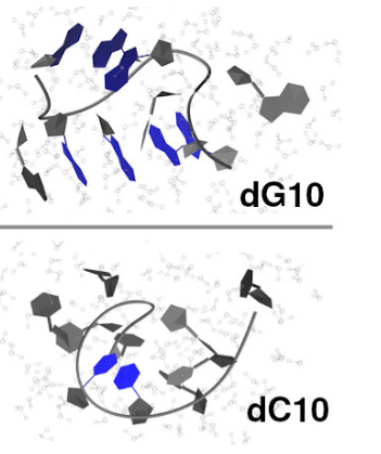
The carbonyl stretching mode is a widely used signal in the infrared spectroscopy of biomolecules, being highly sensitive to changes in their structural organization, such as secondary structure in peptides and proteins or distinct structural motifs in nucleic acids. However, direct mapping between IR spectral bands and specific structural features is not straightforward, particularly for systems with a rugged conformational landscape. Here, we present a computational study of the infrared spectra of C=O stretching modes in DNA bases using a hybrid quantum-classical approach based on the perturbed matrix method. This methodology combines classical molecular dynamics simulations with quantum mechanical calculations on the isolated chromophore, enabling the modeling of vibrational signals across large ensembles of conformations. Recently, we applied the same approach to model the C=O stretching band in thymine-based systems [Parolin et al. PCCP 27.37(2025): 19797-19808]. Here, we expand on that work by investigating guanine- and cytosine-derived systems, thus completing the set of DNA bases containing carbonyl groups and enabling the calculation of C=O stretching in any DNA system. We first benchmark the method on 9-methylguanine and 1-methylcytosine, then extend it to guanine and cytosine 5’-monophosphates, single-stranded deca-homonucleotides, and a seven-base single-stranded DNA oligomer containing all nucleobases. The calculated spectra well reproduce the experimental observations, including frequency shifts and intensity variations arising from stacking and environmental fluctuations, and highlight the role of excitonic effects in shaping spectral features. These results show that the methodology provides insights into how local interactions and conformational dynamics determine the IR spectral signatures of DNA bases.
Download “Article” Modeling_IR_Spectral_Signatures.pdf – Downloaded 1 time – 3 MB
Download a copy of the manuscript